~ The Study of Threes ~
http://threesology.org
A new era in the exploration and teaching of communication must be heralded. In order to do so, we must advance our threesological understanding by imparting it to those whose interest in the field of communication must be forced to acknowledge the persistence and importance of "threes" as a biological, physiological and ideological fact of existence. Without a basice "threes" architecture, communication as we know it would not exist. Thus I implore those of you who wish to participate as part of an avante guard promoting a Revolution which will reverberate into every single subject area, please pass this communiqué on to every instructor of communication.
The form of communication called Speech takes place very difficultly without the mechanism of hearing. Speech is, for the most part, dependent on it. While instructors of speech may well agree that hearing is important, it is not common-place for them to learn or teach about the recurrence of a "threes" pattern in physiology, and how this translates into forms of thought, speech, and/or action. Typically, instructors of communication ideas were not themselves taught to make a psychic connection between a recurring pattern(s) in auditory processes and speaking. Because an important part of communications theory has been overlooked, it is of need that the teachers be sent back to the classroom. For this purpose, let us provide a quick summary for those who are not familiar with the recurrence of "threes" in the structure of the ear, as seen in the image to the left. However, because the research into the threes phenomena has yielded the existence of a developmental physiological trend that can be numerically accessed that appears to follow the same 1, 2, 3 sequence seen in the three Germ layers (Endoderm, Mesoderm, Ectoder), it is necessary to provide an image which shows a perception of the ear whose analysis is said to protray a recurrence of a two-patternd structure (in this case different dichotomies... that is, opposities.)
 Patterns -of- three |
 Patterns -of- two |
In referencing the three Germ layers, the association to a 1, 2, 3 maturational development sequence is identified by the evolutionary condition exhibited by simple organisms (such as sponges appear to) have one germ layer and are called Homoblastic, animals which display a radial symmetry, like cnidarians have two Germ layers and are called Diploblastic, and still more complex organisms (such as humans) which have three Germ layers and are referred to as Triploblastic.
The neural crest cells (NCCs), a transient group of cells that emerges from the dorsal aspect of the neural tube during early vertebrate development has been a fascinating group of cells because of its multipotency, long range migration through embryo and its capacity to generate a prodigious number of differentiated cell types. For these reasons, although derived from the ectoderm, the neural crest (NC) has been called the fourth germ layer....
References:
The Embryo Project Encyclopedia/ Kate McCord
Informatiom about the 3 Germ Layers in Animals/ Komal B. Patil
Neural Crest: The Fourth Germ Layere/ Shyamala K., Yanduri S., Girish HC., Murgod S.
|
| |
| 3 overall divisions: | Outer ear~ Inner ear~ Middle ear |
| 3 middle ear divisions: | Tympanum~ Epitympanum~ Mastoid antrum |
| 3 eardrum membranes: | Cutaneum~ Collagen fibers~ Mucosm |
| 3 semi-circular canals: | Used for balance (equilibrium) |
| 3 bones: | (ossicular chain) Incus~ Stapes~ Malleus |
| 3 main malleus ligaments: | Anterior~ Lateral~ Superior |
| 3 incus anchorage points: | Malleus~ Stapes~ Bony fossa wall |
| 3 cochlea sections: | (Scala) Vestibuli~ Tympani~ Cochlear duct |
| 3 extrinsic muscles (Auricularis): | Anterior~ Superior~ Posterior |
| 3 sound conduction paths: | Electrical~ Mechanical~ Fluid or: Bone (solid)~ Air (gas)~ Fluid (liquid) |
| 3 nerve stimulation paths: | Mechanical~ Chemical~ Electrical |
| 3 outer hair cell rows | typical in mammals but some sources give 3, 4, or 5 |
| Neurotrophin-3 (NT-3) is synthesized by inner and outer hair cells of the developing organ of Corti. | Brain-derived neurotrophic factor (BDNF) is also synthesized. (Prestin is the motor protein of the outer hair cells.) |
| 3 sound qualities: | Pitch~ Volume (intensity)~ Tone |
| 3 sound wave propagation processes: | Diffraction~ Transmission~ Reflection |
| 3 main forms of ossicular chain fixation: | Fluid~ Mechanical~ Otosclerosis |
| 3 classes of ossicular lever action: | Force arm~ Resistance arm~ Fulcrum |
| 3 acoustic distortion forms: | Frequency~ Phase~ Amplitude |
| 3 basic properties of vibrating bodies: | Inertia~ Elasticity~ Dissipation |
| 3 principal types of deafness: | Conduction~ Nerve~Stimulation |
| 3 types of hearing loss: | Conductive~ Sensorineural~ Mixed |
| 3 (inner ear) organs of balance: | Semicircular canals~ Utricle~ Saccule (collectively called the vestibular organ, which can be referred to as {3-in-1} ratio.) |
Source: Language 3's page 1
For some, the consistency of a given pattern(s) from a single source of physiology may be rejected as a coincidence by those who refuse to adopt any other type of mental construct in their conceptualizations. They are not readily able to willingly assess other biological and physiological modalities in order to witness a further recurrence in genetics such as the triplet codings in DNA and RNA, or that humans are on the third planet from a solar energy source, or even give a survey of the following list of threes in human anatomy:
|
http://www.lumen.luc.edu/lumen/MedEd/GrossAnatomy/Threes.html The structure of the human body is organized into groups of three with remarkable frequency. I have listed several examples of this tendency by region. This list does not include structures contributing to form the many "triangles" in the body. If you know of any more, please let me know: --- John A. McNulty, Ph.D. --- mailto:jmcnulty@lumc.edu General 3 layers of skin: Epidermis- Dermis- Hypodermis 3 general venous circulations: Systemic- Pulmonary- Portal Embryology 3 germ layers: Endoderm- Mesoderm- Ectoderm 3 divisions of somites: Sclerotome- Dermatome- Myotome H.O.B. note: At this juncture, let us add a bit of information from the Britannica... The gastrula is the stage of embryonic development at which the embryo appears as three distinct layers of cells (the germ layers): the exterior ectoderm, the middle mesoderm, and the interior endoderm. The mesoderm differentiates to form most of the tissues, structures, and organs of the body. As the embryo lengthens, the mesoderm lying along the midline differentiates to form the notochord, a hollow cartilaginous nerve tube. In the adult the notochord contributes only to the structure of the vertebrae. The mesoderm lateral to this midline then divides into three parts that ultimately form the somites (which subsequently form the vertebrae, the somatic muscles, and the skin), the structures of the urogenital system, and the coelom and its associated structures. (Source: "Muscle." Encyclopædia Britannica Ultimate Reference Suite, 2013.) 3 definitive kidneys: Pronephros- Mesonephros- Metanephros 3 derivatives of skull: Desmo/dermatocranium- Splanchno/viscerocranium- Chondrocranium 3 initial embryonic divisions of the brain: Prosencephalon- Mesencephalon- Rhombencephalon H.O.B. note: At this juncture, let us look at a three-patterned theory concerning the brain: 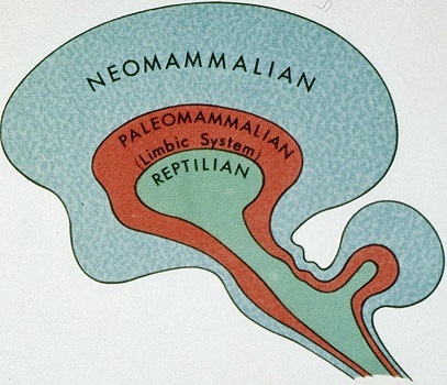 Back 3 parts of the erector spinae m.: Iliocostalis- Longissimus- Spinalis (each are divided again into 3 parts) 3 parts of the transversospinalis m.: Semispinalis m.- Multifidus m.- Rotator m. 3 coverings of the spinal cord: Dura mater- Arachnoid- Pia 3 spaces surrounding spinal cord: Epidural- Subdural- Subarachnoid 3 borders of the scapula: Medial (vertebral)- Lateral- Superior 3 angles of the scapula Neck 3 parts of the axillary artery... 3rd part of axillary artery has 3 branches: Subscapular- Anterior humeral circumflex- Posterior humeral circumflex 3 Brachial plexus trunks: Upper- Middle- Lower 3 posterior Brachial plexus divisions 3 anterior Brachial plexus divisions) 3 Brachial plexus cords: (A) Lateral (B) Medial 3 branches off medial cord: Medial brachial- Medial antebrachial- Medial pectoral (C) Posterior 3 branches off posterior cord: Upper subscapular- Lower subscapular- Thoracodorsal 3 branches off the thyrocervical trunk: Transverse cervical- Suprascapular- Inferior thyroid 3 veins drain the thyroid gland:
3 scalene muscles: Anterior- Middle- Posterior 3 muscles attach to spine of C2:
3 constrictor muscles: Superior- Middle- Inferior 3 parts of the pharynx: Nasopharynx- Oropharynx- Laryngeopharynx 3 parts to the hyoid bone: Body- Greater horn- Lesser horn 3 structures inside carotid sheath: Common carotid artery- Internal jugular vein- Vagus nerve 3 ganglia in sympathetic cervical chain: Superior (magnum)- Middle- Inferior (stellate) Head 3 parts of the maxillary artery: Mandibular- Pterygoid- Pterygopalatine 3 divisions of the trigeminal nerve:
3 cranial fossae: Anterior- Middle- Posterior 3 nuchal (nape of the neck) lines: Iinferior- Superior- Highest 3 layers to calvarium: Outer table- Diploe- Inner table 3 clinoid processes of the sphenoid bone: Anterior- Middle- Posterior 3 cranial nerves (C.N.) pass through jugular foramen: C.N. IX- C.N. X- C.N. XI 3 cranial nerves (C.N.) travel in the wall of the cavernous sinus: C.N. III- C.N. IV- C.N. V 3 cranial nerves (C.N.) carry parasympathetics to the head: C.N. III- C.N. VII- C.N. IX H.O.B. note: At this juncture, let us look at my own three-patterned theory concerning the brain:  3 nasal conchae: Superior- Middle- Inferior 3 principal components of the temporal bone: Squama- Petro/mastoid- Tympanic ring 3 bones of the upper jaw/palate: Premaxilla- Maxilla- Palatine 3 salivary glands: Parotid- Submaxillary- Sublingual 3 muscles innervated by 3 cranial nerves attach to the styloid process:
3 layers in tympanic membrane [eardrum] (derived from 3 germ layers) 3 cranial nerves innervate the external tympanum: C.N. V- C.N. VII- C.N. X 3 middle ear bones: Incus- Malleus- Stapes 3 parts of the ear: Outer- Middle- Inner 3 semicircular canals (of the ear) 3 membranes of the cochlea: Reissner- Tectorial- Basilar 3 intrinsic muscles of the tongue: Longitudinal- Vertical- Transverse 3 muscles elevate the mandible: Masseter m.- Temporalis m.- Medial pterygoid m. 3 branches of the posterior auricular artery:
3 branches of the ophthalmic nerve (VI): Nasociliary n.- Frontal n.- Lacrimal n. 3 superior alveolar nerves:
3 types of deciduous teeth: Incisors- Canine- Molars 3 layers of dentition: Enamal- Dentin- Pulp H.O.B. note: At this juncture, it is of value to note that we humans have a 1st, 2nd and 3rd molar designation as well as the 3rd molars being designated the wisdom teeth. However, it is of notable interest to provide an illustration of the number of teeth in different animals: 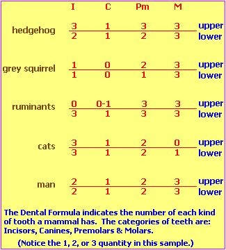 3 general layers of the eye: Retinal- Choroidal/ciliary body- Scleral/corneal 3 color receptors in (eye) cone cells: Red- Blue- Yellow 3 nerves to the larynx:
3 cerebral arteries: Anterior- Middle- Posterior 3 arteries supply the cerebellum:
Shoulder 3 muscles attach to coracoid process: Coracobrachialis- Pectoralis minor- Short head of biceps 3 parts of the axillary artery... (3rd part has 3 branches):
3 muscles of the rotator cuff are rotators 3 glenohumeral ligaments: Superior- Middle- Inferior Arm 3 muscles in anterior comparment: Biceps brachii- Coracobrachialis- Brachialis 3 heads of triceps brachii m.: Long- Lateral- Medial 3 muscles attach to greater tubercle of the humerus: Supraspinatus- Infraspinatus- Teres minor 3 parts of the ulnar collateral ligament of the elbow: Anterior- Posterior- Oblique Forearm 3 muscles in deep anterior compartment:
Hand 3 muscles in thenar eminence:
3 muscles in hypothenar compartment:
3 phalanges in fingers: Proximal- Middle- Distal 3 palmer interossei Thorax 3 compartments: Mediastinum- Left pleural cavity- Right pleural cavity 3 branches off the aortic arch:
3 parts of the aorta: Ascending- Arch- Descending 3 principal surfaces of the pleura: Costal- Diaphragmatic- Mediastinal (the cupola is the superior extension) 3 lobes of the right lung: Superior- Middle- Inferior H.O.B. note: 3 lobes on the right, 2 on the left. 3 (tricuspid) heart valves on the right, 2 (mitral/bicuspid) valves on the left. And thinking in terms of a stroke, where the left hemisphere brain is affected, the right side is affected and vice versa. Attributes of the left brain hemisphere are "three" organized while attributes aligned with the right hemisphere are "two" organized. See the following link for an illustration of this:
3 bronchopulmonary segments of the upper right lobe: Apical- Posterior- Anterior 3 muscles of interthoracic wall:
3 false ribs on each side 3 cusps of the semilunar aortic and pulmonary valves of the heart 3 cusps of the tricuspid (heart) valve 3 main arteries of the heart: Circumflex- Anterior interventricular- Right coronary 3 splanchnic nerves: Greater- Lesser- Least Abdomen 3 folds in anterior abdominal wall:
3 muscles contribute to linea semilunaris/rectus abdominus aponeurosis:
3 openings in diaphragm: Aortic- Esophageal- Inferior vena cava 3 arcuate ligaments of diaphragm:
3 parts of the stomach: Fundus- Body- Pylorus 3 smooth muscle layers of the stomach: Oblique- Longitudinal- Circular 3 unpaired branches of the abdominal aorta:
3 brances of celiac a.: Left gastric a.- Splenic a.- Common hepatic a. 3 brancheds of common hepatic a.: Right gastric a.- Proper hepatic a.- Gastroduodenal a. 3 arterial branches supply adrenal glands: Inferior phrenic- Aorta- Renal 3 structures lie within porta hepatis: Proper hepatic a.- Portal vein- Common bile duct 3 principal veins contribute to the portal vein:
3 parts of the small intestine: Duodenum- Jejunum- Ileum 3 parts of large intestine: Ascending- Transverse- Descending 3 tenia (band-like structure) of large intestine 3 coverings of the spermatic cord:
3 vessels in the umbilical cord: Umbilical vein and 2 umbilical arteries Pelvis and Perineum 3 bones in os coxae: Pubis- Ilium- Ischium 3 parts of the pubis: Body- Superior ramus- Inferior ramus 3 foramina in the pelvis:
3 gluteus muscles: Gluteus maximus- Gluteus medius- Gluteus minimus 3 rectal arteries:
3 branches of the posterior division of internal iliac artery:
3 openings in the urinary bladder: Left ureter- Right ureter- Urethra 3 parts of the urethra: Prostatic- Membranous- Spongy 3 parts of the uterus: Fundus- Body- Cervix 3 erectile compartments of the penis:
3 muscles in superficial perineal pouch:
3 arteries supply the rectum:
3 valves in the rectum: Superior- Middle- Inferior Gluteus 3 gluteal muscles: Gluteus maximus- Gluteus medius- Gluteus minimus 3 muscles innervated by superior gluteal n.:
Thigh 3 hamstring muscles: Biceps femoris- Semitendinosus- Semimembranosus 3 compartments in the femoral sheath 3 muscular compartments of the thigh: Anterior- Medial- Posterior 3 branches of the lateral femoral circumflex a.:
3 muscles contribute to the pes anserinus: Sartorius- Semitendinosus- Gracilis Leg 3 compartments in the leg... (3 muscles in anterior compartment:)
Foot 3 cuneiform bones: Medial- Intermediate- Lateral 3 phalanges in toes: Proximal- Middle- Distal 3 plantar interossei (related to the sole of the foot) Miscellaneous 3 exams in the course http://www.lumen.luc.edu/lumen/MedEd/GrossAnatomy/Threes.html |
H.O.B. note:
Dr. McNulty's comment about the many "Triangles" in the human body is a reference easily passed over by many readers. Some think of a triangle in terms only of a geometric representation of the "three," but it is more than this when be think in terms of what may have influenced the many three-patterned structures in the human body. In so doing, we need to account not only for a pattern-of-three serial event (1 + 1 +1) that could very well develop into a 1 and then 2 and then 3, but one that also exhibits a triangular form that would have impressed this form upon our physiology/anatomy. However, I have also found the existence of examples exhibiting a 1 from 3, 3 into 1, or 3 to 1 (1 to 3) characterization, thus requiring that which influenced (influences) "threes" in our anatomical structure, to also have a triangle and 3 -to- 1 pattern.
Far too many who may encounter this page may be quick to deny that such a pattern recurrence has any value, because they will then offer a handful of examples of some other pattern, thinking that the existence of another pattern other than that being discussed, is a refuation. They will not offer the fact that threesologists do not reject the existence of other patterns, but that the environment of Earth and its immediate external influences, act as a type of buffering system which limits the amount of patterns which are possible. The environment acts as a restraingin mechanism which forces the usage of a 1, 2, 3 (and 3 to 1 ratio) sequencing as a survival mechanism. In other words, the pattern is conserved due to the type(s) of influences environmentally available. The existence of other patterns which can be conceived, attests to the possibility, and not probability of occurrence in our physiology and ideas.
Understanding the issue of a developmental sequence identified numerically, requires an interest in comparative anatomy in order to note differences in "communication". For example, an excerpt from the Britannica on the hearing of differnt animals is revealing in the usage of 3, 2 or 1 auditory structure:
Most lizards can hear. The majority have their best hearing in the range of 400 to 1,500 hertz and possess a tympanum, a tympanic cavity, and a eustachian tube. The tympanum, usually exposed at the surface of the head or at the end of a short open tube, may be covered by scales or may be absent. In general the last two conditions are characteristic of lizards that lead a more or less completely subterranean life. For subterranean lizards airborne sounds are less important than the low-frequency sounds passing through the ground. The middle ear of these burrowers is usually degenerate as well, often lacking the tympanic cavity and eustachian tube. Snakes have neither tympanum nor eustachian tube, and the stapes is attached to the quadrate bone on which the lower jaw swings. Snakes are obviously more sensitive to vibrations in the ground than to airborne sounds. A loud sound above a snake does not elicit any response, provided that the object making the sound does not move or, if it does, the movements are not seen by the snake. On the other hand, the same snake will raise its head slightly and flick its tongue in and out rapidly if the ground behind it is tapped or scratched. Snakes undoubtedly “hear” these vibrations by means of bone conduction. Sound waves travel more rapidly and strongly in solids than in the air and are probably transmitted first to the inner ear of snakes through the lower jaw, which is normally touching the ground, thence to the quadrate bone, and finally to the stapes. Burrowing lizards presumably hear ground vibrations in the same way. All crocodiles have rather keen hearing and have an external ear made up of a short tube closed by a strong valvular flap that ends at the tympanum. The American alligator (Alligator mississippiensis) can hear sounds within a range of 50 to 4,000 hertz. The hearing of crocodiles is involved not only in the detection of prey and enemies but also in their social behaviour; males roar or bellow to either threaten other males or to attract females. Turtles have well-developed middle ears and usually large tympana. Measurements of the impulses of the auditory nerve between the inner ear and the auditory centre of the brain show that the inner ear in several species of turtles is sensitive to airborne sounds in the range of 50 to 2,000 hertz. Source: "Reptile." Encyclopædia Britannica Ultimate Reference Suite, 2013. Note: The article goes on to describe Chemo-reception as a "communications" device as well as an awareness of heat differences. Cleatly then, there are other forms of communication which can be discussed in terms of a 1, 2, 3 maturational development sequence in terms of an appreciation of evolutionary circumstances that some, because of religious inclinations, may prefer to describe as adaptations instead. |
Initial Posting: Tueday, 18-Oct-2016... 12:05 PM
Herb O. Buckland
herbobuckland@hotmail.com